A A A
The size of the font and graphics can be changed as follows: Hold down the CTRL key and press the "+" or "-" key. With CTRL and 0 (zero) you can reset the font size to the standard settings.

Diabetic retinopathy

Diabetic diseases of the eyes could be easily treatable in their early stages

Laser treatments maybe able to help affected persons.

Diabetes mellitus damages many organs, including the eyes, and in particular the retina. In adults from developed countries, diabetic retinopathy is amongst the most common causes of blindness. Treatments such as intravitreal injections or a navigated laser could delay this outcome, or ideally stop it.

Quick facts:

Prevention Regular check-ups by an ophthalmologist, well-managed blood sugar and reducing hypertension are all crucial.

Treatment The most common treatment for retinal swelling is injections into the eye. This can be combined with laser treatment for leaking vessels that targets many individual spots in the field of view.

Symptoms Black spots (“floaters”), red haze, blurred and fuzzy image, flashes of light, dark curtain in the field of view.

Technical terms Diabetes mellitus, diabetic retinopathy, diabetic maculopathy, diabetic macular edema (DME), proliferative retinopathy, non-proliferative retinopathy.

    Quick Facts Netzhauterkrankungen

    Diabetic retinopathy – effects

    Fortgeschrittenes Stadium Diabetische Retinopathie

    Vision with advanced stage diabetic retinopathy

    Frühes Stadium Diabetische Retinopathie

    Vision with early stage diabetic retinopathy

    Gesundes Auge

    Vision with a healthy eye

      In diabetes, patients often have high concentrations of sugar in their blood. This damages smaller vessels, resulting in circulation disorders. It also stimulates the formation of new vessels. These are of very poor quality and brittle, which can lead to bleeding in the eye. If leaking fluid accumulates at the site of sharpest vision (the macula), visual acuity will deteriorate. Scars form, and eyesight is threatened.

      Stages of diabetic retinopathy

      A distinction is made between early and advanced forms of diabetic retinopathy. There are significant differences in the clinical picture. The same is true for treatment options.

      Non-proliferative retinopathy

      Nicht-proliferative Retinopathie

      In this case, the patient has a mild form of diabetic retinopathy (early stage). Slight visual impairment may occur, but this won’t necessarily be the case.

      The cause: Elevated blood glucose levels lead to fats and proteins being stored in the vascular walls and layers of nerve fibers. These hard exudates develop over a long time.

      Laser treatment, for example with the Navilas® Laser System, can be helpful in severe cases of non-proliferative diabetic retinopathy.

      Proliferative retinopathy

      Proliferative Retinopathie

      This is a severe form of diabetic retinopathy (late stage). It threatens the fundamental functioning of the eye.

      In diabetes, the body responds to the insufficient supply of oxygen and nutrients. This is why it forms new, substandard blood vessels. If bleeding then occurs, this can drastically deteriorate vision. This can happen very quickly. If the body’s own healing process then causes the substandard vessels in the eye to scar, the retina can detach. In the worst-case scenario, this can lead to blindness or loss of the eye.

      Diabetic macular edema

      Both forms of diabetic retinopathy may be accompanied by edema. This is where fluid builds up in the macula. The macula is the part where vision is sharpest. The eye can be considerably and permanently affected by this condition. 

      Diabetic retinopathy – late onset of symptoms

      At the early stage of retinopathy, the patient initially has no symptoms at all. Only an ophthalmologist will be able to discover the first signs on the retina. Symptoms such as foggy or unclear vision or dark spots in the field of view are indications that the disease has progressed.

      Symptoms

      • Black spots (“floaters”)
      • Red haze
      • Blurred and fuzzy image
      • Flashes of light
      • Dark curtain in the field of view

      The diagnosis – as quickly as possible

      Every diabetes patient must undergo regular ophthalmological examinations. Even when the results are normal: frequent check-ups by the ophthalmologist are essential. This is because retinopathy can only be treated promptly when it is detected early. Important: Well-managed blood glucose and blood pressure could help avoid disease progression.

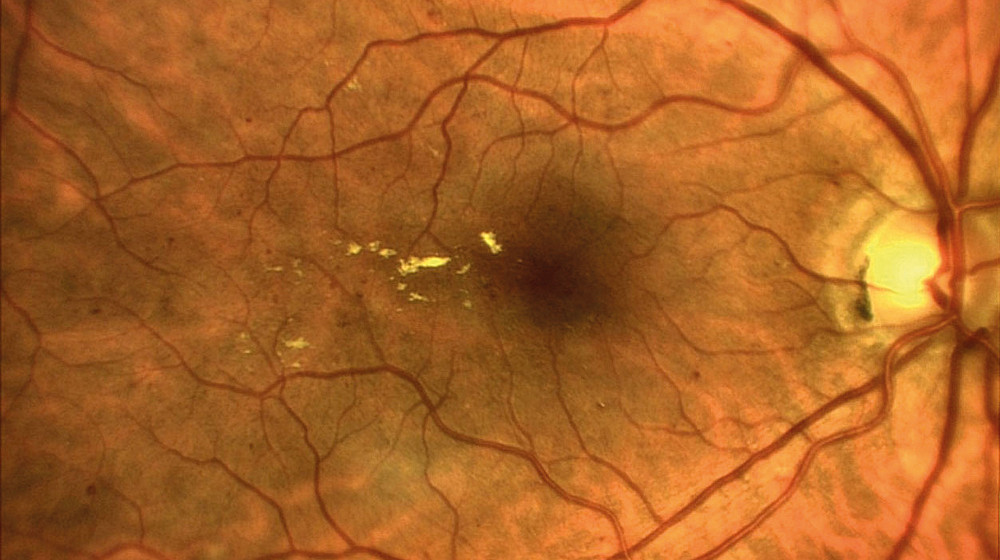
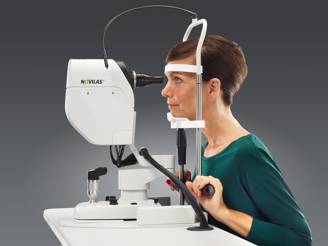
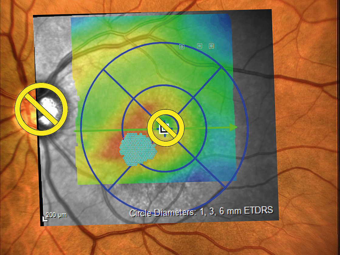
      The examination: Changes to the retina are visible on the slit lamp. If these are present, this is followed by further diagnostic steps. One of these is fluorescein angiography. In this test, a dye is injected via a vein in the arm, which allows the vessels in the eye to be seen. Several photos are taken during the examination. These are used to assess whether and where the laser has to be used. "Optical coherence tomography" (OCT) is then used for a more precise examination. It depicts the individual layers of the retina and allows a number of changes to be detected. This makes it possible to determine the stage of diabetic retinopathy more accurately. It is thus easier for the doctor to define a treatment strategy for the patient.

      OCT

      OCT cross section through the macula

      OCT Dickenkarte

      OCT thickness map

      Fluorescein angiography

      Spaltlampe

      Slit lamp

        Recording of focal laser therapy

        Treating diabetic retinopathy with lasers

        There are different approaches to the treatment of diabetic retinopathy. Affected persons can receive helpful treatment at each stage of the disease depending on the changes they have experienced. The greatest prerequisite for success: correct and consistent treatment for the underlying disease, diabetes mellitus.

        Find out more about treatment with the Navilas® Laser System

        Intravitreal injection

        Intravitreal injection

        If there is already swelling of the retina, the injection of medications can help. A specific antibody against a messenger substance produced by the body is injected into the center of the eye. This antibody inhibits the growth of new blood vessels. Studies have shown that this can lead to a significant reduction in edema. However, these injections have to be repeated regularly, often for life. To avoid infections that can lead to blindness, these injections in the eye have to be carried out under strict hygiene conditions. To monitor the effect of the injections, OCT examinations should be carried out regularly (often on a monthly basis) to determine the thickness of the retina.

        Intravitreale Injektion - shutterstock
        Injektion in das Auge - shutterstock

        Laser treatment for swelling

        Laser treatment for swelling

        A laser is often used in addition to an injection. It can help to make the effect of the injections last longer. The laser targets defective blood vessels or insufficiently supplied areas and obliterates them. The Navilas® Laser System provides ophthalmologists with particularly good assistance in this regard. The precise localization of the diseased area by the doctor, supported by computer-assisted planning in the case of Navilas, increases the effect. Before starting treatment, all of the examination images are overlaid by the computer. The ophthalmologist only marks the spots that need to be targeted. Unlike with conventional lasers, the Navilas® Laser System helps to hit these exact spots marked by the doctor.

        Even in patients who respond poorly or not at all to injection therapy (around 30% of macular edema patients), your ophthalmologist may consider laser treatment for the swelling to prevent the disease from advancing further.

        Learn more about Navilas

        Treatment example of focal laser coagulation

        Tissue-sparing, microsecond pulsing therapy laser

        Tissue-sparing, microsecond pulsing therapy laser

        There is a new type of laser available for use, especially in the early stages of the disease. Initial studies have shown it to be effective and tissue-sparing. In contrast to normal laser treatment, where the laser output is applied continuously, microsecond pulsing therapy applies the laser in a number of short bursts. It is believed that obliteration does not take place, thus, there is no permanent damage to the photoreceptors caused by the laser. However, the stimulation of the tissue can still achieve improvements in swelling and decrease edema. To do so, the area of the swelling must first be carefully identified and planned with individual points. Full coverage is required for the treatment to achieve its intended effect. Navilas® is an excellent aid to doctors here, because in using the tissue-protecting application, a doctor with a conventional laser has no information about areas that have already been treated. With digital pre-planning and documentation from the Navilas® Laser System, however, this information is available and transparent. Insufficient coverage can be avoided.

        Learn more about Navilas

        Stimulation of tissue using microsecond pulsing therapy

        Peripheral laser treatment

        Peripheral laser treatment

        If new diseased vessels become visible, there is another treatment to help. In this procedure, many individual points in the patient’s outer field of view are delivered with the laser. This allows the central area of the retina to receive a better supply of oxygen. The Navilas® laser also proves to be a valuable assistant in this area, as an even distribution of the laser points through pre-planning helps to obtain successful treatment results. The pattern function – in which multiple points are applied in a row – and the pre-planning function of the Navilas® laser both enable the treatment to be performed quickly.

        Learn more about Navilas

        Navigation-supported, peripheral treatment

        Creating and executing the digital treatment plan

        “My many years of experience with Navilas® show that it is possible to reduce the number of injections and achieve more stable treatment results. This results in less burden on our patients.”

        Prof. Dr. Marcus Kernt, Munich

        We would like to point out that OD-OS, as a device manufacturer, does not answer any questions about individual disease pattern, does not make any diagnoses or perform examinations or treatments. The present website is a pure knowledge platform, which is the result of direct communication with physicians and serves as a source of information for you. Whether or not the therapies described are suitable for you can only be decided by your ophthalmologist - on the basis of a comprehensive examination of your state of health and your eyes. Please also carefully read the „Legal Notice“ and our „Privacy Policy“.

        Do you have questions?

        Contact us

        Welcome at Navilas for Patients

        Are you a physician?

        Physician website

        or

        Are you a patient?

        Continue